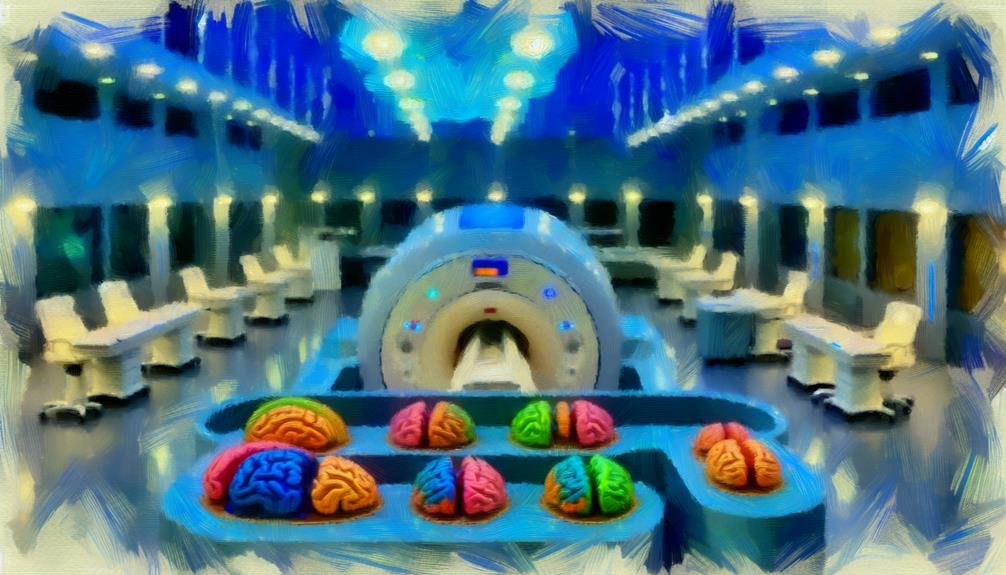
Functional magnetic resonance imaging (fMRI) is a powerful tool for studying brain activity, offering advantages such as non-invasive measurement, high spatial resolution, and real-time data collection. These features enable researchers to observe brain function dynamically and apply findings across diverse populations. Nevertheless, fMRI also has notable limitations. Its temporal resolution is slower than neuronal firing, and data can be influenced by motion artifacts and physiological noise. Additionally, high costs and limited facility availability can restrict access. Misinterpretations of fMRI data can further complicate research outcomes. Exploring these pros and cons reveals critical understanding into fMRI's role in neuroscience.
Main Points
- fMRI provides non-invasive measurement of brain activity, allowing for safer long-term studies of brain dynamics without surgical risks.
- It offers high spatial resolution, enabling precise localization of brain functions, crucial for understanding complex cognitive processes.
- Real-time data collection in fMRI allows for observation of dynamic brain activity during specific tasks, supporting applications like neurofeedback.
- However, fMRI has limited temporal resolution, making it difficult to accurately capture the timing of neural activities.
- The high cost and limited availability of fMRI facilities restrict accessibility and can delay diagnosis and treatment for patients.
Advantages of Fmri
Functional magnetic resonance imaging (fMRI) is a powerful neuroimaging technique that offers several remarkable advantages in the field of cognitive neuroscience and clinical research. One of its primary benefits is the ability to non-invasively measure brain activity by detecting changes in blood flow, which correlates with neuronal activity. This allows researchers to identify specific brain regions involved in various cognitive processes, enhancing our understanding of brain function.
Another important advantage of fMRI is its high spatial resolution, enabling detailed mapping of brain structures and networks. This precision is essential for both theoretical studies and clinical applications, such as pre-surgical planning for patients with brain tumors or epilepsy.
Additionally, fMRI can be used to investigate the brain's response to stimuli in real-time, making it a vital tool for studying dynamic processes such as perception, decision-making, and motor control.
Furthermore, fMRI is a versatile technique applicable across diverse populations, including healthy individuals and those with neurological disorders. Its ability to provide observations into both typical and atypical brain function contributes to the development of targeted interventions and therapies, ultimately advancing our knowledge of the human brain.
Non-Invasive Brain Imaging
Non-invasive brain imaging techniques have revolutionized the way researchers and clinicians study brain structure and function. These methods, which include functional magnetic resonance imaging (fMRI), positron emission tomography (PET), and electroencephalography (EEG), allow for the examination of neural activity without the need for surgical intervention. This non-invasive nature not only minimizes risk to patients but also promotes the longitudinal study of brain dynamics across various conditions and interventions.
The ability to visualize brain activity in real time has greatly advanced our understanding of neuropsychological disorders, cognitive processes, and the impact of therapeutic approaches. For example, fMRI provides revelations into the hemodynamic response associated with neuronal activation, thereby enabling clinicians to identify functional abnormalities related to conditions such as epilepsy, stroke, and psychiatric disorders.
Additionally, non-invasive imaging techniques are instrumental in research settings, allowing for the exploration of brain-behavior relationships while preserving participant comfort.
On the other hand, while these methods provide important information, they are not without limitations, including concerns about spatial and temporal resolution, as well as the interpretation of complex data.
Nevertheless, the benefits of non-invasive imaging in enhancing our comprehension of the human brain are undeniable.
Real-Time Data Collection
Real-time data collection is a crucial aspect of functional magnetic resonance imaging (fMRI), enabling researchers to capture dynamic brain activity as it occurs. This capability allows for the observation of neural processes in action, facilitating a deeper understanding of cognitive functions, emotional responses, and sensory perceptions. By acquiring data in real-time, fMRI can provide immediate feedback regarding brain activation patterns associated with specific tasks, enhancing the potential for immediate adjustments in experimental design.
One notable advantage of real-time fMRI is its application in neurofeedback, where individuals can learn to regulate their brain activity through visual or auditory feedback. This technique shows promise for treating various psychological conditions, such as anxiety or depression, by allowing patients to engage in self-modulation of their brain states.
However, challenges exist, including the need for sophisticated algorithms and processing techniques to handle the extensive amount of data generated. Additionally, the real-time nature of the method may introduce noise and artifacts that can complicate interpretation.
Despite these hurdles, the potential of real-time data collection in fMRI remains a powerful tool for advancing our understanding of the human brain and its complex functions.
High Spatial Resolution
A key advantage of fMRI is its high spatial resolution, which allows for precise localization of brain activity. This feature enables researchers to differentiate between activity in adjacent brain regions, improving the understanding of functional anatomy. High spatial resolution is particularly important in studies investigating complex cognitive functions, such as language, memory, and decision-making.
The ability to observe fine-grained details in brain activation patterns contributes to the identification of region-specific functions, eventually advancing neuroscience. Below is a comparison illustrating the spatial resolution of fMRI relative to other neuroimaging techniques:
| Neuroimaging Technique | Spatial Resolution (mm) |
|---|---|
| fMRI | 1-3 |
| PET | 4-6 |
| EEG | 10+ |
As depicted in the table, fMRI's superior spatial resolution makes it a preferred choice for studies requiring detailed mapping of brain activity. This capability not only improves the accuracy of findings but also supports the exploration of complex neural networks, opening the door for deeper understanding into brain function.
Limitations of Fmri
While fMRI offers extraordinary viewpoints into brain activity, it is not without its limitations. One notable constraint is its reliance on hemodynamic responses, which are indirect indicators of neural activity. This leads to a temporal resolution that is considerably slower than the actual neuronal firing, making it challenging to capture rapid cognitive processes accurately.
Additionally, fMRI is sensitive to various confounding factors, such as motion artifacts and physiological noise from heartbeats and breathing. These variables can obscure the data and complicate the interpretation of results. Furthermore, the method typically requires participants to remain still, which can be difficult for certain populations, such as children or individuals with motor impairments.
Another limitation involves the complexity of data analysis. fMRI data is often high-dimensional and requires sophisticated statistical methods for interpretation, which can introduce biases or errors if not handled correctly.
Moreover, the results of fMRI studies can sometimes lack ecological validity, as laboratory settings may not accurately reflect practical cognitive processes.
Cost and Accessibility Issues
The implementation of functional magnetic resonance imaging (fMRI) is hindered by considerable equipment costs, which can limit its adoption in various healthcare settings.
Additionally, the limited availability of fMRI machines in certain geographic locations further exacerbates accessibility issues for patients in need of this advanced neuroimaging technique.
As a result, these financial and logistical barriers may restrict the potential benefits of fMRI in clinical and research environments.
High Equipment Costs
Facing considerable financial barriers, the implementation of functional Magnetic Resonance Imaging (fMRI) technology is often limited by its high equipment costs. The initial investment required to purchase fMRI machines can range from several million dollars to upwards of ten million, depending on the model and capabilities. This substantial expense not only includes the cost of the machine itself but also necessitates additional funding for installation, maintenance, and operational expenses.
The high costs can restrict the number of facilities that can afford to offer fMRI services, ultimately affecting research and clinical practices. The table below highlights key financial aspects related to fMRI equipment:
| Aspect | Estimated Cost |
|---|---|
| Equipment Purchase | $2 million – $10 million |
| Installation | $500,000 – $1 million |
| Maintenance (annual) | $100,000 – $200,000 |
| Operational Expenses | Variable (staff, utilities) |
These financial considerations underscore the challenges faced by institutions seeking to utilize fMRI technology, limiting its accessibility and potentially hindering advancements in neuroscience and medical diagnostics.
Limited Availability Locations
Accessibility remains a considerable concern in the domain of functional Magnetic Resonance Imaging (fMRI), as the limited availability of scanning facilities poses challenges for both researchers and patients. This scarcity is particularly pronounced in rural and underserved urban areas, where advanced medical imaging technologies are often lacking. As a result, individuals requiring fMRI services may face notable travel burdens, which can lead to delays in diagnosis and treatment.
Moreover, the concentration of fMRI facilities in major academic and medical centers can exacerbate inequities in access to care. Patients may encounter long waiting times for appointments, further complicating their healthcare journeys. This limited access not only affects patient outcomes but also hinders the progress of research studies that rely on diverse participant populations to improve the generalizability of findings.
In addition to geographical disparities, the high operational costs associated with fMRI technology often necessitate prioritization of certain research projects and clinical applications over others. This prioritization can restrict the exploration of less common conditions or groundbreaking research avenues, eventually stalling advancements in understanding brain function and pathology.
Thus, addressing the limited availability of fMRI facilities is vital for equitable healthcare and scientific progress.
Potential for Misinterpretation
The interpretation of fMRI data can lead to considerable misunderstandings regarding brain activity, particularly when the complexities of neural responses are oversimplified.
Additionally, the contextual factors surrounding an experiment can heavily influence the results, potentially skewing conclusions drawn from the data.
As a result, a careful evaluation of both the methodology and the context is essential to avoid misinterpretation.
Brain Activity Misunderstandings
Misinterpretations of brain activity data derived from fMRI can lead to considerable misunderstandings regarding cognitive processes and emotional states. The complexity of neural activity often results in oversimplified conclusions about behavior and thought patterns. For instance, detecting heightened activity in specific brain regions does not necessarily equate to a direct correlation with particular cognitive functions. This can promote misconceptions about the brain's role in decision-making, emotional regulation, or even social interactions.
Furthermore, the brain operates through elaborate networks rather than isolated regions, and fMRI primarily captures regional blood flow changes, which may not accurately reflect underlying neural mechanisms. Consequently, claiming that activation in a specific area signifies a definitive cognitive process can mislead interpretations of psychological phenomena.
Additionally, the temporal resolution of fMRI is limited, making it challenging to associate specific brain activity with precise moments in time. This can obscure the dynamic nature of cognitive functions, leading to erroneous assumptions about the timing and sequence of brain events.
In the end, such misunderstandings can affect research outcomes, clinical practices, and public perceptions of neuroscience, underscoring the need for careful interpretation of fMRI data within the broader context of cognitive neuroscience.
Contextual Influence on Results
Context plays an essential role in shaping the outcomes of fMRI studies, often leading to misinterpretations of the results. The neural correlates identified in such studies can vary markedly based on the experimental design, participant characteristics, and environmental factors. Researchers must exercise caution when generalizing findings, as the contextual nuances can dramatically alter the interpretation of brain activity.
Several factors contribute to the potential for misinterpretation in fMRI research:
- Task-Specific Variability: Different cognitive tasks can activate overlapping brain regions, making it challenging to pinpoint specific functions.
- Participant Demographics: Variations in age, gender, and cultural background can influence neural responses and skew results.
- Environmental Context: The setting in which fMRI scans are conducted, including the presence of stressors or distractions, may affect participant performance and brain activity.
Understanding these contextual influences is critical for accurately interpreting fMRI data. Researchers should endeavor to account for these variables to improve the reliability and validity of their findings, thereby minimizing the risk of drawing misleading conclusions from fMRI studies.
Common Questions
How Does Fmri Compare to Other Brain Imaging Techniques?
fMRI excels in evaluating brain activity through blood flow changes, providing high spatial resolution. Comparatively, techniques like PET offer metabolic understandings, while EEG captures temporal dynamics, each serving distinct purposes in neuroscience research and clinical applications.
Can Fmri Be Used for Clinical Diagnosis?
Functional magnetic resonance imaging (fMRI) has potential in clinical diagnosis, particularly for neurological disorders. Nevertheless, its application is often supplementary to established diagnostic methods, emphasizing the importance of an all-encompassing approach to patient evaluation and management.
What Safety Precautions Are Necessary During an Fmri Scan?
Safety precautions during an fMRI scan include screening for metallic implants, ensuring the patient is free from ferromagnetic objects, monitoring essential signs, and providing ear protection to minimize discomfort from scanner noise.
How Is Patient Comfort Maintained During Fmri Procedures?
Patient comfort during fMRI procedures is maintained through the use of cushioning, adjustable headrests, noise-canceling headphones, and clear communication about the procedure, along with providing adequate ventilation and minimizing scan duration whenever possible.
What Future Advancements Are Expected in Fmri Technology?
Future advancements in fMRI technology are anticipated to include improved imaging resolution, real-time data processing, integration with artificial intelligence for better analysis, and the development of portable instruments for broader clinical and research applications.
Conclusion
In conclusion, functional magnetic resonance imaging (fMRI) presents considerable advantages, including its non-invasive nature, real-time data collection capabilities, and high spatial resolution, which aid thorough brain studies. Nevertheless, limitations such as high costs and accessibility issues, along with the potential for misinterpretation of results, must be acknowledged. A balanced understanding of these pros and cons is essential for effectively utilizing fMRI in both clinical and research settings, ensuring accurate observations into brain function and activity.

